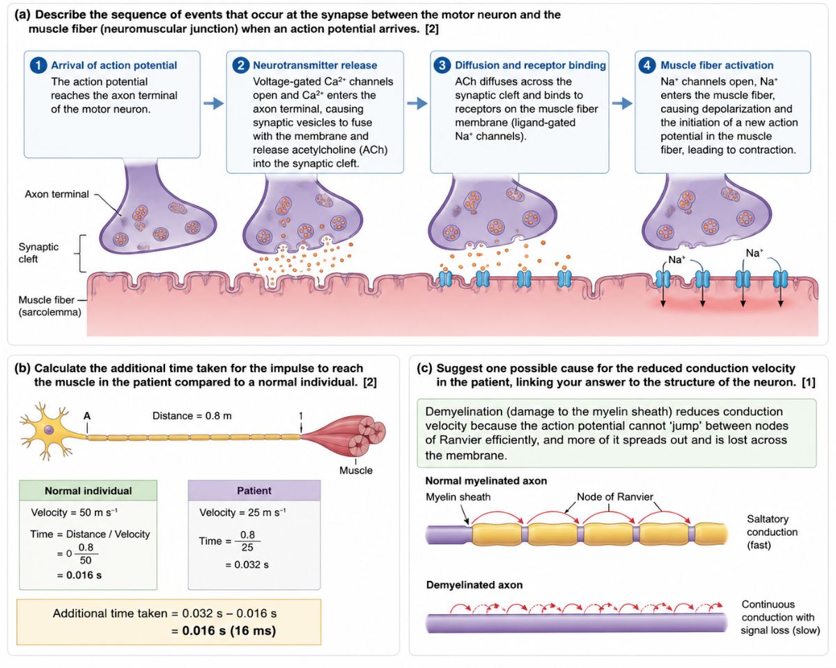
From electrical signal to muscle contraction — in milliseconds.
Have you ever thought about what actually happens between the moment your brain sends the command "lift your arm" and the moment your arm actually moves? The gap between your motor neuron and your muscle fibre is where the magic — and the science — truly happens. This junction, called the neuromuscular junction (NMJ), is one of the most elegant signalling systems in the human body.
Let's break it down, step by step.
What Is the Neuromuscular Junction?
The neuromuscular junction is the synapse — a specialised communication point — between the terminal end of a motor neuron and a muscle fibre. Unlike the electrical signals that travel along nerves, communication across this junction is chemical. A signal must be converted from electrical to chemical and back again, all within a fraction of a second.
Step-by-Step: What Happens When an Action Potential Arrives
Step 1 — Arrival of the Action Potential
The journey begins in the motor cortex of the brain, travels down the spinal cord, and races along the motor neuron until it reaches the axon terminal — the very tip of the nerve. At this point, the electrical signal (the action potential) has arrived at the boundary between neuron and muscle.
Step 2 — Neurotransmitter Release
The arrival of the action potential triggers voltage-gated calcium (Ca²⁺) channels to open in the axon terminal membrane. Calcium ions flood into the terminal, and this is the critical trigger: the calcium causes synaptic vesicles — tiny membrane-bound sacs packed with the neurotransmitter acetylcholine (ACh) — to fuse with the axon terminal membrane and release their contents into the synaptic cleft, the narrow gap between neuron and muscle.
Think of this step like a postal worker being handed a key (calcium) that allows them to open the warehouse (vesicles) and release the packages (ACh) into the gap.
Step 3 — Diffusion and Receptor Binding
Acetylcholine molecules diffuse rapidly across the synaptic cleft — a gap of roughly 20–50 nanometres — and bind to ligand-gated Na⁺ (sodium) channels embedded in the muscle fibre membrane (the sarcolemma). These receptors are highly specific; only ACh can bind to them and trigger their opening.
Step 4 — Muscle Fibre Activation
Once ACh binds to its receptors, the ligand-gated sodium channels open. Sodium ions (Na⁺) rush into the muscle fibre down their concentration gradient, causing depolarisation of the sarcolemma. This depolarisation initiates a new action potential in the muscle fibre itself, which spreads along the membrane and deep into the fibre through structures called T-tubules — ultimately triggering muscle contraction.
The signal has been received. The muscle moves.
The Speed of the Signal: How Fast Does It All Happen?
Here's where it gets fascinating from a physics perspective. Nerve impulses don't all travel at the same speed — and the difference matters enormously.
In a healthy individual, a motor neuron might conduct an impulse at 50 m s⁻¹. Over a distance of 0.8 metres (from a nerve point to a muscle), that means the signal arrives in just:
Time = Distance ÷ Velocity = 0.8 ÷ 50 = 0.016 s (16 ms)
Now consider a patient whose conduction velocity has been reduced to 25 m s⁻¹:
Time = 0.8 ÷ 25 = 0.032 s (32 ms)
The additional time taken in the patient compared to the healthy individual is:
0.032 − 0.016 = 0.016 s (16 ms)
Sixteen milliseconds might sound trivially small, but in neurology, this kind of delay is clinically significant. It can mean slower reflexes, muscle weakness, or impaired coordination — and it points directly to a structural problem with the neuron itself.
Why Would Conduction Be Slower? Demyelination Explained
The most common reason for reduced nerve conduction velocity is demyelination — damage to or loss of the myelin sheath.
What Is the Myelin Sheath?
Myelin is a fatty insulating layer wrapped around the axons of many neurons by specialised cells (Schwann cells in the peripheral nervous system). It doesn't cover the axon continuously — it leaves small gaps called nodes of Ranvier exposed at regular intervals.
Saltatory Conduction — Nature's Fast Lane
In a healthy myelinated axon, the action potential doesn't travel as a continuous wave along the entire membrane. Instead, it "jumps" from one node of Ranvier to the next — a process called saltatory conduction (from the Latin saltare, to leap). This is dramatically faster and more energy-efficient than continuous conduction.
What Happens When Myelin Is Damaged?
When the myelin sheath is damaged — as occurs in conditions like multiple sclerosis (MS), Guillain-Barré syndrome, or as a result of certain autoimmune diseases — the action potential can no longer jump efficiently between nodes. Instead, it must spread continuously and slowly across the exposed membrane, losing amplitude as it goes. The result is:
Slower conduction velocity
Signal degradation over longer distances
In severe cases, complete conduction block
This is why demyelinating diseases are so debilitating: the neurons themselves may be intact, but the insulation that makes fast signalling possible has been compromised.
Why This Matters: Clinical Relevance
Understanding the neuromuscular junction and nerve conduction isn't just academic — it underpins diagnosis and treatment of a wide range of conditions:
Myasthenia Gravis — an autoimmune disease where antibodies attack ACh receptors at the NMJ, preventing the signal from reaching the muscle effectively. The result is progressive muscle weakness that worsens with use.
Multiple Sclerosis — a demyelinating disease of the central nervous system causing slowed or blocked conduction, leading to a range of motor and sensory symptoms.
Nerve Conduction Studies (NCS) — clinicians measure the speed at which impulses travel along nerves to detect demyelination, diagnose neuropathies, and monitor disease progression. Exactly the kind of calculation shown in Part (b) of the question above.
Botulinum Toxin (Botox) — works precisely at the NMJ by blocking the release of ACh from synaptic vesicles, preventing muscle contraction. Used medically for spasticity, and cosmetically to reduce wrinkles.
Key Vocabulary Summary
Term Definition Neuromuscular junction (NMJ) Synapse between a motor neuron and a muscle fibre Axon terminal The end of a motor neuron where neurotransmitter is released Synaptic cleft The narrow gap between neuron and muscle fibre Acetylcholine (ACh) Neurotransmitter released at the NMJ Sarcolemma The cell membrane of a muscle fibre Saltatory conduction Action potential "jumping" between nodes of Ranvier Myelin sheath Fatty insulating layer that speeds conduction Node of Ranvier Gaps in the myelin sheath where depolarisation occurs Demyelination Damage or loss of the myelin sheath, slowing conduction
Final Thought
The neuromuscular junction is a masterpiece of biological engineering — converting an electrical signal into a chemical message, releasing it across a nanometre-scale gap, binding it to exactly the right receptor, and firing off a new electrical signal, all in under a millisecond. When this system works, we barely notice it. When it doesn't — through demyelination, autoimmune attack, or toxin exposure — the consequences range from mild weakness to complete paralysis.
Next time you pick up a cup of coffee, remember: that simple motion involved thousands of neuromuscular junctions firing in perfect coordination, each one executing this four-step chemical relay at speeds you can barely imagine.
This blog is part of the INACADEMICS IB Biology and IGCSE Biology revision series. For more exam-focused content on the nervous system, muscle physiology, and beyond, visit inacademics.com.